The Sternal Angle
 |
| http://www.freeclipart.pw/uploads/2017/05/tags-halloween-skeletons -halloween-did-you-know-halloween-skeletons--17.jpeg |
The sternal angle is also called the Angle
of Louis or the manubriosternal joint. It is found on the sternum between the
manubrium (the upper part of the sternum) and the 10cm long sternal body as a
transverse ridge.
 |
| http://anatomyzone.com/wp-content/uploads/2015/02/sternal-angle.png |
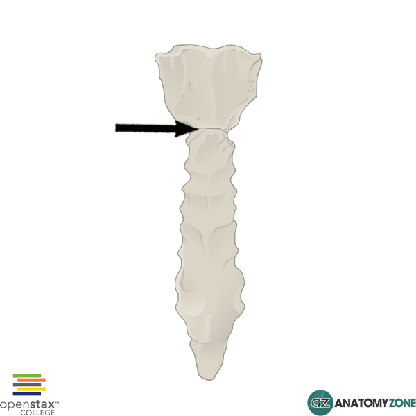
If you need any help identifying the sternal angle,
it can be palpated 5cm below the suprasternal notch, where the
sternal body meets the manubrium at an angle of 15 degrees.
Joint type: the sternal angle is
a secondary cartilaginous joint, also known as a symphysis joint. (https://clinanat.com/mtd/282-sternal-angle-of-louis).
This basically means it is made up of a plate of fibrocartilage connected to
the articular hyaline surfaces of the bone.
 |
| http://teachmeanatomy.info/wp-content/uploads/Great-vessels1.jpg |
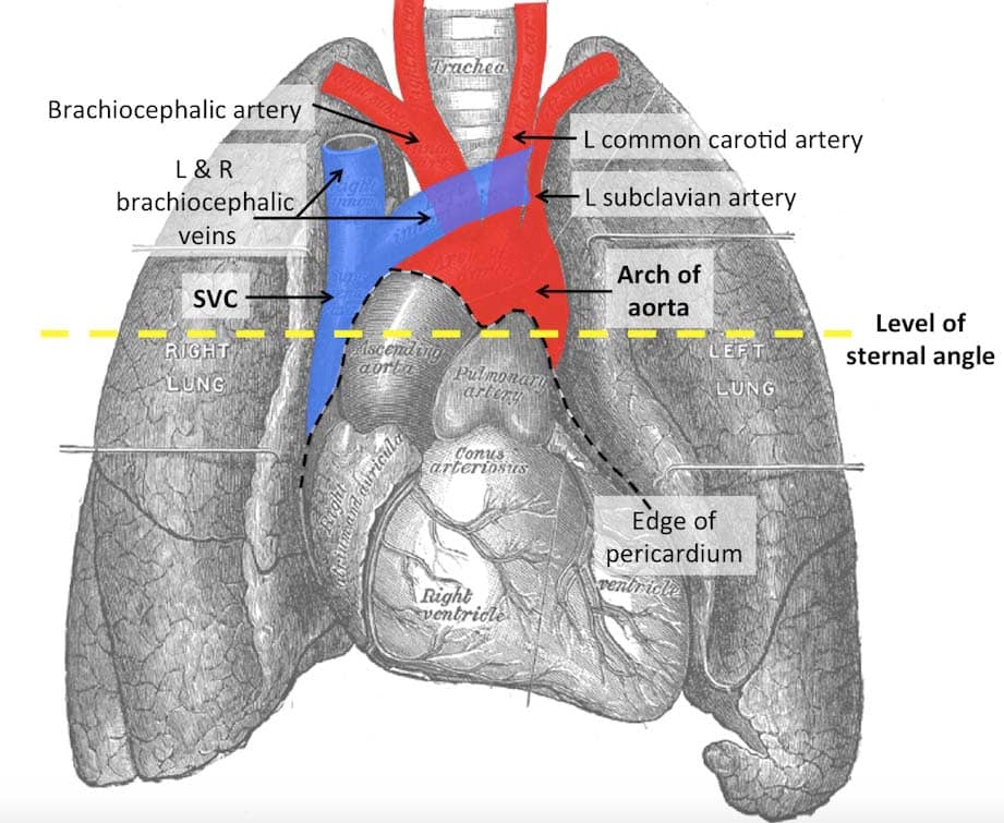
Relations:
The sternal angle is a useful landmark in the
thorax, as it can be used as a reference point for key anatomical structures in the area. An easy way to remember the important structures that passes behind the sternal angle is the mneumonic: RAT PLLANT. N.B. this mnemonic does not describe the structures in order of appearance, and is just to remember the structures in no particular order.
 |
| RAT PLLANT |
R = rib (the second rib)
A = aortic arch – the beginning and end of the
aortic arch is found on this plane
T = tracheal bifurcation – where the trachea
divides into left and right
P = Pulmonary trunk
L = ligamentum arteriosum
L = left recurrent laryngeal
A = azygos vein
N = nerves
T = thoracic duct
Surface anatomy: The
Angle of Louis is found 5cm below suprasternal notch
 |
| https://www.intechopen.com/source/html/18257/media/image3.jpeg |
The angle is at the level of the second costal
cartilage
It is found between T4 and T5 vertebra posteriorly
(http://medchrome.com/basic-science/anatomy/sternal-angle-of-louis-significance-of-landmark/)
- the body of the sternum is opposite T5 to T8.
Clinical relevance:
•
- The
second rib is found at the level of the thoracic plane, this can be used as a
reference point when finding the valves of the heart.
•
- If the
left recurrent laryngeal is damaged the patient will present with a hoarse
voice
•
- The
sternal angle separates the mediastinum into superior and inferior parts – an
important point to note is that the terminology is slightly different in
radiology, and to not get confused by this in clinics
--> The
Superior Mediastinum extends from the thoracic inlet to T4
--> The Posterior Mediastinum extends from T4 to the diaphragm, an
 |
| http://thegoofyanatomist.weebly.com/uploads/3/0/9/9/30995885/3218989_orig.jpg |
--> The sternum is normally protected from injury by the presence of the elastic costal cartilages, but it may be fractured with dislocation of the thoracic spine.
Interesting fact from the sternal angle lecture
You can get a stab wound through bone – this
surprised me, but was used as an example in the sternal angle lecture to
demonstrate what structures lie behind the sternal angle, and could be at risk
if you get stabbed at this point, for example, the arch of the aorta, the
azygos system etc. We then talked about the consequences of a stab wound
through bone for a while in more general terms, and the complications that can
arise from a bone stab wound. These include…
- Osteomyelitis
 |
| https://www.wpclipart.com/cartoon/assorted/more_cartoons/bone_cartoon.png |
- Shattered bone (in the humerus this could impinge
on nerves such as the radial nerve)
- Bone necrosis (- by impinging on blood supply, as
is the case for the scaphoid bone
- Fat embolism
History:
The sternal angle was named after the French
physician, Antoine Louis
References:
http://anatomyzone.com/tutorials/musculoskeletal/sternal-angle/
Gray's Anatomy
Teaching from the Anatomy Summer School
Rat Image - https://s-media-cache-ak0.pinimg.com/originals/2d/1f/b8/2d1fb82a184c5b72d4a04f36277dd82b.jpg


Comments
Post a Comment