Coeliac Trunk: Step by Step
Today: The Abdominal Aorta Part 1 the Coeliac Trunk
Recently I have been busy trying to get my head around the abdominal aorta - the largest vessel in the abdomen, and the one artery that supplies all of the abdominal organs. So without further ado, a few introductions are in order.

Introducing.....the Abdominal Aorta!
So if you want to meet the abdominal aorta, where should you look? Well the best place to spot it at the beginning of its journey is with its friends - the azygos vein and thoracic duct. BUT watch out - it's incredibly shy, and likes hiding away at the back of the abdomen (in the retroperitoneal space).
All three friends go into the abdomen together through a small opening called the aortic opening of the diaphragm at T12.
It's a bit of a "pushover" and is shifted to the left of the abdomen by the INFERIOR VENA CAVA (so it can be found posterior and to the left of the inferior vena cava)on top of the lumbar vertebrae.
 First: The Coeliac Trunk
The first major branch of the abdominal aorta is the coeliac trunk. It supplies the foregut, basically the lower oesophagus, stomach, liver, spleen pancreas and proximal duodenum
It is found at T12 and splits into three branches:
1. Common hepatic artery
2. Splenic artery
3. Left gastric artery
This can be remembered with the acronym Left Hand Side.
Let's look at each of these in a bit more detail...
1. The common hepatic:
This eventually supplies the liver and bile duct as the proper hepatic, but first let's trace its journey from the coeliac trunk....
- The common hepatic artery moves away from the coeliac trunk to the right of the body.
- As it moves, it gives off a branch called the gastroduodenal artery.
- Once it has given off the gastroduodenal artery branch, the common hepatic artery continues as the proper hepatic artery.
So what happens next?
The proper hepatic artery continues to the liver, where it divides into the right and left hepatic arteries to supply different areas of the liver.
- Once the common hepatic artery reaches the liver, it splits into the right and left hepatic arteries. The right hepatic artery gives off a branch known as the cystic artery which will supply the gall bladder.
But earlier on in the journey, before the proper hepatic artery reaches the liver, it gives off a branch to the stomach. This is called the right gastric artery.

I know it's a lot to take in, so make sure you pause and understand this bit before reading any further - this is kind of a skeleton structure for everything that is yet to come with the common hepatic artery. Everything else I write about will build on what we have just covered. To help check you get it, I found some pictures online to demonstrate the simple structure and the key branches. See how many you can identify before moving on...
Problem 1: Label the branches off the common hepatic artery on the diagram below... (a--> f)

Problem 2: label the abdominal aorta branches on the diagram below (a-->g)
Problem 3: look at the radiogram. It shows the celiac trunk coming off from the abdominal aorta and the three branches. We are only looking at the common hepatic branch labels (a-->d). Can you label them
Questions:
5. At what level is the coeliac trunk?
6. The coeliac trunk is to the ****** of the Inferior vena cava?
7. How many main branches are there off the coeliac trunk?
Answers:
1. a = right hepatic, b = cystic, c = proper hepatic, d = gastroduodenal, e = right gastric, f = left hepatic , g = common hepatic
2. a = right hepatic, b = left hepatic artery, c = gastroduodenal artery, d = common hepatic artery, e = right gastric artery f = abdominal aorta, g = splenic artery, h = left gastric artery
3. a = right hepatic artery, b = left hepatic artery, c = gastroduodenal, d = common hepatic artery
4. a = right hepatic artet, b = left hepatic artery, c = common hepatic artery, d = left gastric artery, e = gastroduodenal arty, f splenic artery
5. Coeliac trunk = T12
6. Left
7. Three: left gastric, common hepatic, splenic.
 The branches of the common hepatic and proper hepatic: a bit more on the right gastric artery and the gastroduodenal
So now you understand the basic structure of the common hepatic artery, let's look at its two main branches - the right gastric artery and the gastroduodenal.
a. The right gastric - this is the easiest branch to understand as it simply supplies the lesser curvature (the small c) of the stomach.
b. The gastroduodenal artery - this is slightly more complicated.
It gives off one branch: the right gastroepiploic - but in some textbooks this is referred to as the right gastro-omental. Why? Well if you split the name up - gastro = stomach, omental = omentum. This refers to the greater omentum, which is kind of like an "apron" attached to the greater curvature of the stomach (just below the stomach, it is also attached to the transverse colon)...
What does it do?
Well the greater omentum plays a key role in the immune system. It is made up of fat, blood vessels, lymph and tissue, and has three major functions:
1. Fat storage - the greater omentum is fattier in obese patients than thinner patients where it is more translucent.
2. Removes cell debris - through the presence of white blood cells
3. Isolates infections and trauma
( see more: https://www.reference.com/science/function-omentum-708766e94ff9d5e2#)
.
The omentum has three basic functions. It stores fat deposits, it contains milky spots that contain white blood cells, which boost immunity by getting rid of cellular debris, and it isolates wounds and infections by wrapping around infected areas
It is important you remember the right gastric artery from the previous section and the right gastro-omental artery that we have just mentioned, because we are not finished with them yet...they will make a reappearance later on in the coeliac trunk, but for now just remember where they originate from. All shall become clear :)
- After giving off the right-gastro-omental, the gastroduodenal continues as the superior pancreaticoduodenal.

Superior pancreaticoduodenal - the complicated bit.....
Don't worry - we have nearly finished with the common hepatic branches now, and soon you will be a master of it. But before we can move on and review everything we need to just finish off with the gastroduodenal.
So we know that it has come off from the common hepatic and given off the right gastro-omental artery - right? Well now it continutes as the superior pancreaticoduodenal artery to supply the pancreas.
In the next picture I am going to get rid of most of the diagram and just show you the gastroduodenal branch so it is easier to imagine....

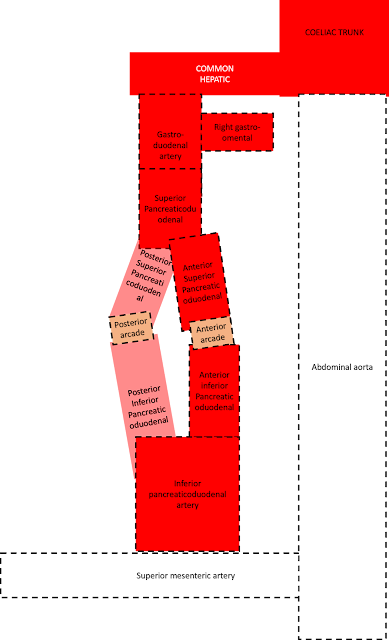
So after continuing as the superior pancreaticoduodenal, the artery decides to split into two branches. One to supply the front (anterior) of the pancreas, and one to supply the behind (the posterior part of the pancreas).
Therefore these vessels are called - you guessed it - the posterior superior pancreaticoduodenal and the anterior superior pancreaticoduodenal.

 As these go down behind and in front of the pancreas, they join with the posterior inferior pancreaticoduodenal arteries and the anterior inferior pancreaticoduodenal arteries at the bottom of the pancreas. We say that they connect through posterior and anterior arterial arcades.
Once the anterior and posterior inferior pancreaticoduodenal arteries are formed, they join together after supplying the pancreas to form the inferior pancreaticoduodenal arteries.
SO where do the inferior pancreaticoduodenal arteries go? Well this is the neat thing - they actually come off from a branch of the abdominal aorta, a bit further down than the coeliac trunk. This branch is called the superior mesenteric artery. I will add it into the diagram with some dotted lines so you can see exactly what I mean...
You might have heard of the superior mesenteric artery before, and it is an extremely important branch off the abdominal aorta - so much so that I will probably dedicate a whole blog post to it, but for now, it's just enough to know that one branch off the superior mesenteric is the inferior pancreaticoduodenal which links with all the arteries we have just talked about.
Does that make sense? Are you ready for a review?
Just before we add some questions in to check our understanding, I just wanted to add in one final detail. A small branch off the anterior superior pancreaticoduodenal which will link to an artery we haven't yet met....but more on that later.
Problem 1: I like these reviews because you get a chance to see the vessels in context with their organs. I will go through the exact relations between the vessels and their organs later, but it's nice to get an idea as we go alongSo let's start off with the stomach....
a. = gastrosuodenal
b = superior pancreaticoduodenal
c. = right gastro-omental
d. = hepatic arteries
e. = coeliac trunk
2. a. = gastroduodenal
b. = anterior and posterior superior pancreaticouodenal
c superior mesenteric artery
d= post and anterior inferior pancreaticoduodenal
3. a = right gastro-omental
b. = gastroduodenal
c. - posterior superior pancreaticoduodenal
d = anterior superior pancreaticoduodenal
e. = anterior inferior pancreaticoduodenal
f. posterior inferior pancreaticoduodenal g = superior mesenteric artery
4 a = left gastric
b = common hepatic
c = right gastric
d = gastroduoenal
e. = anterior suoerior pancreaticoduodenal
f = right gastro-omental
5. a. = proper hepatic
b = gastroduodenal
c. = right gastric
d = superior pancreaticoduodenal
e. = inferior pancreaticoduodenal
f = superior mesenteric
g = left gastric
h = splenic
I = common hepatic
6. Answer = inferior pancreaticoduodenal
7. Answer = E = superior mesenteric
2. The left Gastric Artery
So now onto something a bit easier - the left gastric. Can you remember the right gastric artery from earlier? Well the left gastric artery is basically its counterpart on the left hand side, except instead of arising from the common hepatic artery, it comes straight from the coeliac trunk and supplies the lesser curvature (the small c) of the stomach.
On the way it gives a branch to the oesophagus (the oesophageal artery) and actually connects to the right gastric artery on the lesser curvature of the stomach.
3, The Splenic artery
Finally we are almost at the end of our journey - we have reached the splenic artery. Compared to the other branches of the coeliac trunk, this follows a much more windy course to its destination - the spleen. It kind of looks like a snake.
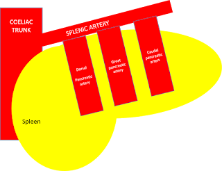
The first major branch it gives off is the dorsal pancreatic artery to the pancreas. This is followed by two further branches to the pancreas: the great pancreatic artery and the caudal pancreatic artery.
 These arteries all join the inferior pancreatic artery (also known as the transverse pancreatic artery).
- The dorsal pancreatic artery in particular, is special, because it joins to the anterior superior pancreaticoduodenal - remember the mystery artery from before? Yep, that was the dorsal pancreatic artery - so this is another anastomosis, providing a connection between the common hepatic branch of the coeliac trun and the splenic artery of the coeliac trunk. It might be easier if I show this on a picture....
Later the splenic artery gives a branch to form the left gastro-omental (also known as left gastro-epiploic), which is the counterpart to the right gastro-omental and supplies the greater curvature of the stomach
SO....that's it folks: the final branch of the coeliac trunk done and dusted. Just a few questions to finish up with, but then that's the Coeliac trunk done. Stick around for the superior mesenteric artery because I think that will probably be next as we journey down the abdominal aorta. Thanks for sticking with me :)
1. What supplies the foregut?
A. The Inferior mesenteric artery
B. The superior mesenteric artery
C. The Coeliac trunk
D. The common hepatic
E. The left gastric
Answer = C = the coeliac trunk
2. Which artery supplies the fundus of the stomach?
A. RIght gastric
B. Left gastric
C. Right epiploic
D. Left epiploic
E. Short gastric
Answer = E = short gastric
3. If the coeliac trunk was blocked, would the stomach still receive blood?
Answer = yes, remember the connection from the superior mesenteric artery? The stomach receives collaterals through this system.
Fact of the day: Never squeeze a boil on your cheek....it could result in 6th nerve palsy! This is because the veins in the face drain to a structure known as the cavernous sinus. This is basically a space in the head containing the nerves III, IV, V and Vi. If pus is sxqueezed into the vein draining into this sinus, an abscess can form, placing pressure on the sixth nerve!
Recently I have been busy trying to get my head around the abdominal aorta - the largest vessel in the abdomen, and the one artery that supplies all of the abdominal organs. So without further ado, a few introductions are in order.

Introducing.....the Abdominal Aorta!
So if you want to meet the abdominal aorta, where should you look? Well the best place to spot it at the beginning of its journey is with its friends - the azygos vein and thoracic duct. BUT watch out - it's incredibly shy, and likes hiding away at the back of the abdomen (in the retroperitoneal space).
All three friends go into the abdomen together through a small opening called the aortic opening of the diaphragm at T12.
It's a bit of a "pushover" and is shifted to the left of the abdomen by the INFERIOR VENA CAVA (so it can be found posterior and to the left of the inferior vena cava)on top of the lumbar vertebrae.
 First: The Coeliac Trunk
The first major branch of the abdominal aorta is the coeliac trunk. It supplies the foregut, basically the lower oesophagus, stomach, liver, spleen pancreas and proximal duodenum
It is found at T12 and splits into three branches:
1. Common hepatic artery
2. Splenic artery
3. Left gastric artery
This can be remembered with the acronym Left Hand Side.
Let's look at each of these in a bit more detail...
1. The common hepatic:
This eventually supplies the liver and bile duct as the proper hepatic, but first let's trace its journey from the coeliac trunk....
- The common hepatic artery moves away from the coeliac trunk to the right of the body.
- As it moves, it gives off a branch called the gastroduodenal artery.
- Once it has given off the gastroduodenal artery branch, the common hepatic artery continues as the proper hepatic artery.
So what happens next?
The proper hepatic artery continues to the liver, where it divides into the right and left hepatic arteries to supply different areas of the liver.
- Once the common hepatic artery reaches the liver, it splits into the right and left hepatic arteries. The right hepatic artery gives off a branch known as the cystic artery which will supply the gall bladder.
But earlier on in the journey, before the proper hepatic artery reaches the liver, it gives off a branch to the stomach. This is called the right gastric artery.

I know it's a lot to take in, so make sure you pause and understand this bit before reading any further - this is kind of a skeleton structure for everything that is yet to come with the common hepatic artery. Everything else I write about will build on what we have just covered. To help check you get it, I found some pictures online to demonstrate the simple structure and the key branches. See how many you can identify before moving on...
Problem 1: Label the branches off the common hepatic artery on the diagram below... (a--> f)

Problem 2: label the abdominal aorta branches on the diagram below (a-->g)
Problem 3: look at the radiogram. It shows the celiac trunk coming off from the abdominal aorta and the three branches. We are only looking at the common hepatic branch labels (a-->d). Can you label them
Problem 4: This is more difficult - it's a picture from a dissection of the common hepatic arter. Ignore all the labels that are not lettered and see if you can figure out what features a --> e are pointing to.
Questions:
5. At what level is the coeliac trunk?
6. The coeliac trunk is to the ****** of the Inferior vena cava?
7. How many main branches are there off the coeliac trunk?
Answers:
1. a = right hepatic, b = cystic, c = proper hepatic, d = gastroduodenal, e = right gastric, f = left hepatic , g = common hepatic
2. a = right hepatic, b = left hepatic artery, c = gastroduodenal artery, d = common hepatic artery, e = right gastric artery f = abdominal aorta, g = splenic artery, h = left gastric artery
3. a = right hepatic artery, b = left hepatic artery, c = gastroduodenal, d = common hepatic artery
4. a = right hepatic artet, b = left hepatic artery, c = common hepatic artery, d = left gastric artery, e = gastroduodenal arty, f splenic artery
5. Coeliac trunk = T12
6. Left
7. Three: left gastric, common hepatic, splenic.
 The branches of the common hepatic and proper hepatic: a bit more on the right gastric artery and the gastroduodenal
So now you understand the basic structure of the common hepatic artery, let's look at its two main branches - the right gastric artery and the gastroduodenal.
a. The right gastric - this is the easiest branch to understand as it simply supplies the lesser curvature (the small c) of the stomach.
b. The gastroduodenal artery - this is slightly more complicated.
It gives off one branch: the right gastroepiploic - but in some textbooks this is referred to as the right gastro-omental. Why? Well if you split the name up - gastro = stomach, omental = omentum. This refers to the greater omentum, which is kind of like an "apron" attached to the greater curvature of the stomach (just below the stomach, it is also attached to the transverse colon)...
What does it do?
Well the greater omentum plays a key role in the immune system. It is made up of fat, blood vessels, lymph and tissue, and has three major functions:
1. Fat storage - the greater omentum is fattier in obese patients than thinner patients where it is more translucent.
2. Removes cell debris - through the presence of white blood cells
3. Isolates infections and trauma
( see more: https://www.reference.com/science/function-omentum-708766e94ff9d5e2#)
.
The omentum has three basic functions. It stores fat deposits, it contains milky spots that contain white blood cells, which boost immunity by getting rid of cellular debris, and it isolates wounds and infections by wrapping around infected areas
It is important you remember the right gastric artery from the previous section and the right gastro-omental artery that we have just mentioned, because we are not finished with them yet...they will make a reappearance later on in the coeliac trunk, but for now just remember where they originate from. All shall become clear :)
- After giving off the right-gastro-omental, the gastroduodenal continues as the superior pancreaticoduodenal.

Superior pancreaticoduodenal - the complicated bit.....
Don't worry - we have nearly finished with the common hepatic branches now, and soon you will be a master of it. But before we can move on and review everything we need to just finish off with the gastroduodenal.
So we know that it has come off from the common hepatic and given off the right gastro-omental artery - right? Well now it continutes as the superior pancreaticoduodenal artery to supply the pancreas.
In the next picture I am going to get rid of most of the diagram and just show you the gastroduodenal branch so it is easier to imagine....

So after continuing as the superior pancreaticoduodenal, the artery decides to split into two branches. One to supply the front (anterior) of the pancreas, and one to supply the behind (the posterior part of the pancreas).
Therefore these vessels are called - you guessed it - the posterior superior pancreaticoduodenal and the anterior superior pancreaticoduodenal.

 As these go down behind and in front of the pancreas, they join with the posterior inferior pancreaticoduodenal arteries and the anterior inferior pancreaticoduodenal arteries at the bottom of the pancreas. We say that they connect through posterior and anterior arterial arcades.
Once the anterior and posterior inferior pancreaticoduodenal arteries are formed, they join together after supplying the pancreas to form the inferior pancreaticoduodenal arteries.
SO where do the inferior pancreaticoduodenal arteries go? Well this is the neat thing - they actually come off from a branch of the abdominal aorta, a bit further down than the coeliac trunk. This branch is called the superior mesenteric artery. I will add it into the diagram with some dotted lines so you can see exactly what I mean...
I took off the other branches of the coeliac trunk just to make the diagram clearer - but what do you think? Isn't that amazing?
In Anatomy when two vessels join like this it is called an "anastomosis" or more formally (according to Wikipedia!): a cross-connection between adjacent channels, tubes, fibres, or other parts of a network
You might have heard of the superior mesenteric artery before, and it is an extremely important branch off the abdominal aorta - so much so that I will probably dedicate a whole blog post to it, but for now, it's just enough to know that one branch off the superior mesenteric is the inferior pancreaticoduodenal which links with all the arteries we have just talked about.
Does that make sense? Are you ready for a review?
Just before we add some questions in to check our understanding, I just wanted to add in one final detail. A small branch off the anterior superior pancreaticoduodenal which will link to an artery we haven't yet met....but more on that later.
Problem 1: I like these reviews because you get a chance to see the vessels in context with their organs. I will go through the exact relations between the vessels and their organs later, but it's nice to get an idea as we go alongSo let's start off with the stomach....
Problem 2:
Problem 3:
Problem 4: let's start combining everything we have looked at so far. First the stomach...
Problem 5:
6. The superior pancreaticoduodenal artery anastomoses with?
A. The Splenic
B. Common Hepatic
C. Right Gastric
D. Inferior pancreaticoduodenal
E. Oesophageal
7. The inferior pancreaticoduodenal branches from which vessel?
A. Coeliac trunk
B. Splenic
C. Common hepatic
D. Inferior Mesenteric
E. Superior mesenteric
1. a. = gastrosuodenal
b = superior pancreaticoduodenal
c. = right gastro-omental
d. = hepatic arteries
e. = coeliac trunk
2. a. = gastroduodenal
b. = anterior and posterior superior pancreaticouodenal
c superior mesenteric artery
d= post and anterior inferior pancreaticoduodenal
3. a = right gastro-omental
b. = gastroduodenal
c. - posterior superior pancreaticoduodenal
d = anterior superior pancreaticoduodenal
e. = anterior inferior pancreaticoduodenal
f. posterior inferior pancreaticoduodenal g = superior mesenteric artery
4 a = left gastric
b = common hepatic
c = right gastric
d = gastroduoenal
e. = anterior suoerior pancreaticoduodenal
f = right gastro-omental
5. a. = proper hepatic
b = gastroduodenal
c. = right gastric
d = superior pancreaticoduodenal
e. = inferior pancreaticoduodenal
f = superior mesenteric
g = left gastric
h = splenic
I = common hepatic
6. Answer = inferior pancreaticoduodenal
7. Answer = E = superior mesenteric
2. The left Gastric Artery
So now onto something a bit easier - the left gastric. Can you remember the right gastric artery from earlier? Well the left gastric artery is basically its counterpart on the left hand side, except instead of arising from the common hepatic artery, it comes straight from the coeliac trunk and supplies the lesser curvature (the small c) of the stomach.
On the way it gives a branch to the oesophagus (the oesophageal artery) and actually connects to the right gastric artery on the lesser curvature of the stomach.
 |
| Sorry - mislabelled, spleen should read pancreas |
Finally we are almost at the end of our journey - we have reached the splenic artery. Compared to the other branches of the coeliac trunk, this follows a much more windy course to its destination - the spleen. It kind of looks like a snake.
The first major branch it gives off is the dorsal pancreatic artery to the pancreas. This is followed by two further branches to the pancreas: the great pancreatic artery and the caudal pancreatic artery.
 These arteries all join the inferior pancreatic artery (also known as the transverse pancreatic artery).
 - The dorsal pancreatic artery in particular, is special, because it joins to the anterior superior pancreaticoduodenal - remember the mystery artery from before? Yep, that was the dorsal pancreatic artery - so this is another anastomosis, providing a connection between the common hepatic branch of the coeliac trun and the splenic artery of the coeliac trunk. It might be easier if I show this on a picture....
Later the splenic artery gives a branch to form the left gastro-omental (also known as left gastro-epiploic), which is the counterpart to the right gastro-omental and supplies the greater curvature of the stomach
Finally, the splenic arterty ends by sending branches to the stomach (short gastric) which supplies the fundus of the stomach and to the spleen (the splenic branches).
SO....that's it folks: the final branch of the coeliac trunk done and dusted. Just a few questions to finish up with, but then that's the Coeliac trunk done. Stick around for the superior mesenteric artery because I think that will probably be next as we journey down the abdominal aorta. Thanks for sticking with me :)
1. What supplies the foregut?
A. The Inferior mesenteric artery
B. The superior mesenteric artery
C. The Coeliac trunk
D. The common hepatic
E. The left gastric
Answer = C = the coeliac trunk
2. Which artery supplies the fundus of the stomach?
A. RIght gastric
B. Left gastric
C. Right epiploic
D. Left epiploic
E. Short gastric
Answer = E = short gastric
3. If the coeliac trunk was blocked, would the stomach still receive blood?
Answer = yes, remember the connection from the superior mesenteric artery? The stomach receives collaterals through this system.
Fact of the day: Never squeeze a boil on your cheek....it could result in 6th nerve palsy! This is because the veins in the face drain to a structure known as the cavernous sinus. This is basically a space in the head containing the nerves III, IV, V and Vi. If pus is sxqueezed into the vein draining into this sinus, an abscess can form, placing pressure on the sixth nerve!


Comments
Post a Comment